RESEARCH/R&D
 Advances in machine learning and artificial intelligence techniques offer a promise to supplement rapid, accurate, and reliable computer-assisted disease screening. Such techniques are particularly valuable in overburdened and/or resource constrained regions. These regions also tend to exhibit high prevalence of infectious diseases and report high mortality. Our research in machine learning and artificial intelligence algorithms aims to improve disease detection accuracy and reliability, with a goal to also explain algorithm behavior.
Tuberculosis (TB) is one of the world’s deadliest communicable diseases and widespread, particularly in resource-challenged parts of the world. The Director-General of the World Health Organization (WHO) reports that TB kills five thousand people every day! TB is also severe comorbidity in immunocompromised HIV+ patients and is correlated with poor control in developing nations. According to WHO report , over 900,000 people were living with HIV/TB in 2017, with the disease having claimed 300,000 lives that year.
Since the lack of sufficient radiological services in the area suggests the utility of automation to perform the screening, our research aims to apply our expertise in image analysis and machine learning toward developing novel computational solutions to analyze chest x-rays (CXR) and screen for diseases. Radiological analysis of CXR images is a routine component in diagnosing pulmonary TB.
Our research includes developing machine learning algorithms, deep learning, in particular, to perform image classification, detection, and localization tasks. Deep learning algorithms offer high scalability and the potential to deliver superior results with end-to-end feature extraction and classification. These algorithms analyze images for feature that are consistent with radiological manifestations of pulmonary diseases. We also develop novel deep model ensembles to improve classification reliability and visualization of machine decisions toward explaining the inner-workings of the algorithm.
To test our work, we are collaborating with the Indiana University School of Medicine, and AMPATH (Academic Model Providing Access to Healthcare), an NGO supported by USAID that runs the largest AIDS treatment program in sub-Saharan Africa, and Moi Teaching and Referral Hospital (MTRH), in Eldoret Kenya. AMPATH has designed and developed a mobile radiology vehicle with lightweight digital x-ray units that are readily transportable in rural areas. Their staff takes CXRs of the population and screens them, using software containing our algorithms, for the presence of disease.
Additional Links:
NLM InFocus
SPIE Newsroom
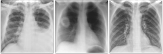
CXR images showing 2 examples of pulmonary abnormalities (left: pleural effusion, middle: cavitary lung lesion right lung), and normal lung image (right).
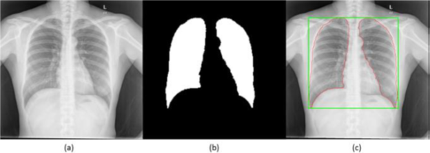
PA Chest X-ray lung ROI segmentation process: (a) original image, (b) computed lung mask, (c) segmented lung ROI with the bounding box.
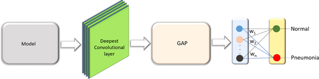
Customized architecture of pretrained models.
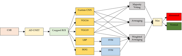
Ensemble learning for CXR classification
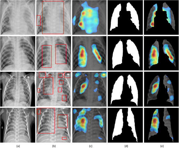
Visual explanations through gradient-based localization using CAM: (a) Input CXRs; (b) Bounding boxes localizing regions of activations; (c) CAM showing heat maps superimposed on the original CXRs; (d) Automatically segmented lung masks; (e) CAM showing heat maps superimposed on the cropped lungs. images showing 2 examples of pulmonary abnormalities (left: pleural effusion, middle: cavitary lung lesion right lung), and normal lung image (right).